
Dog's Third Eyelid Explained: Normal vs Abnormal by Vet
A dog's third eyelid refers to the pinkish to whitish membrane positioned in the inner corner of each eye, serving essential roles in safeguarding and moisturizing the eyeball. Integrative veterinarian Dr. Julie Buzby provides a detailed overview of the third eyelid's structure and offers guidance o
A dog's third eyelid refers to the pinkish to whitish membrane positioned in the inner corner of each eye, serving essential roles in safeguarding and moisturizing the eyeball. Integrative veterinarian Dr. Julie Buzby provides a detailed overview of the third eyelid's structure and offers guidance on appropriate actions when this membrane protrudes unusually. Furthermore, she delves into seven distinct issues that can affect the third eyelid in dogs.
While humans rely solely on their upper and lower eyelids to shield the eyes from particles, dust, foreign objects, and intense illumination, dogs and cats possess an additional protective feature known as the third eyelid, providing an extra barrier of defense.
Typically, in healthy conditions, this third eyelid remains neatly concealed and barely noticeable. Nevertheless, specific circumstances might cause it to rise or exhibit irregularities, which could point to an underlying health concern that warrants attention.
What Exactly is the Third Eyelid in Dogs?
The third eyelid, also called the nictitating membrane, consists of a slender, pinkish-white layer situated beneath the lower eyelid in the nasal corner of the eye. Its primary functions encompass several vital tasks, including:
- Gliding horizontally over the eyeball to offer supplementary shielding from dust particles and potential scratches
- Clearing away debris that accumulates on the eye's surface
- Spreading the tear film evenly to ensure the eye remains properly lubricated
- Containing the gland of the third eyelid, which is a key producer of tears
One can liken the third eyelid to a combination of a windshield wiper or squeegee mechanism and a deployable protective shield for the eye. Moreover, it incorporates a built-in reservoir of tear fluid, akin to windshield washer solution, to support eye health.
Understanding the Anatomy of the Third Eyelid
The structural design of the third eyelid enables it to perform these critical protective duties effectively. Both its inner and outer layers are lined with conjunctiva, the same delicate pinkish-white tissue that covers the insides of the upper and lower eyelids and extends onto the eyeball's surface. In numerous dogs, though not universally, a darker pigmented margin appears along the leading edge where it is most visible.
Nestled within this conjunctival covering is a T-shaped cartilaginous structure that imparts the necessary firmness, allowing the membrane to sweep away debris and tears across the eye's surface. This cartilage functions much like an integrated squeegee, with the wiping edge positioned at the forefront of the third eyelid and the supportive stem reaching back toward the eye's inner corner.
Positioned discreetly beneath the third eyelid at the base of this cartilage's handle is the gland responsible for the third eyelid. This gland significantly contributes between 30% and 60% of the tear fluid, underscoring its pivotal role in maintaining ocular moisture levels.
Is the Third Eyelid Visible in Dogs?
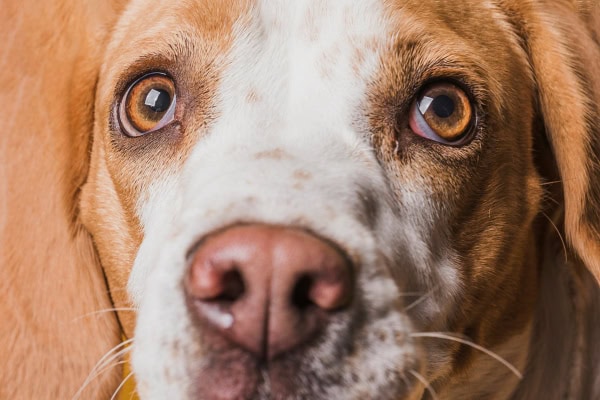
In a healthy, alert dog's eye, the third eyelid appears only as a thin, flat sliver of pinkish to dark tissue hugging the inner margin of the eyeball. Without close inspection, it might go entirely unnoticed.
Certain breeds like Pugs, Boxers, English Bulldogs, French Bulldogs, and Boston Terriers often display more conspicuous third eyelids due to their facial structure. In contrast, for many other breeds, spotting it requires deliberate effort.
If you regularly observe your dog's eyes, you likely have a baseline understanding of how much of the third eyelid is typically visible under normal conditions. Establishing this norm is crucial for identifying any deviations that might signal trouble.
Situations Where Seeing More of the Third Eyelid is Normal
Certain scenarios can naturally cause a greater portion of the third eyelid to become apparent without indicating pathology. For instance, a larger segment might emerge when the dog is in a relaxed state or asleep. Brief elevations can also occur during periods of stress, excitement, or rapid eye movements.
Additionally, the third eyelid may extend further across the eye surface during activities necessitating heightened protection, such as navigating dusty environments or brushing through vegetation.
Should You Worry if Your Dog's Third Eyelid is Prominent?
Generally speaking, if the third eyelid becomes more visible than your dog's usual baseline—and this occurs outside of contexts like sleep, excitement, or protective needs—it may suggest an irregularity. This concern heightens if the protrusion persists over time or accompanies other clinical signs.
Recognizing Symptoms of Third Eyelid Disorders
Canine third eyelid disorders manifest through diverse symptoms influenced by the specific impact on the eye. Among the most frequently observed are:
- A vivid pink or red fleshy protrusion located in the inner eye corner
- Greater than normal coverage of the eyeball by the third eyelid
- Appearance of redness, bumps, or swelling on the third eyelid
- Excessive tearing leading to dampened fur surrounding the eye
- Unusual discharge appearing yellow or green, potentially watery or thick and mucous-like
- Crusty buildup of dried secretions around the eyes
- Redness or puffiness affecting the upper and lower eyelid tissues
- Squinting or keeping the eye shut owing to discomfort and swelling
- Frequent pawing at the eyes, signaling irritation and pain
When to Schedule a Veterinary Visit for Your Dog
Immediate veterinary consultation is essential if the third eyelid is elevated alongside redness, pain, or cloudiness in the eye. Even in milder presentations without overt distress, persistent elevation merits professional evaluation.
The urgency and nature of symptoms will dictate whether a standard appointment or emergency care is advised. Ocular conditions can progress rapidly, so prompt assessment is advisable to prevent complications.
What Happens During a Veterinary Examination?
For suspected third eyelid concerns, the veterinarian initiates a thorough physical assessment. Diagnosis may stem from this exam alone, though supplementary diagnostics are common to pinpoint the issue accurately. These can include:
Detailed Ophthalmic Examination
Employing focused illumination, the vet inspects pupil size and light reactivity. A handheld ophthalmoscope further scrutinizes the eye's exterior and interior, revealing potential inflammation, neurological involvement, or other anomalies influencing the third eyelid.
Fluorescein Dye Application
To detect corneal ulcers associated with third eyelid elevation, fluorescein staining is applied. This dye adheres to damaged corneal areas, crucial since irritation might lead to self-inflicted scratches or ulcers from rubbing.
Tear Production Assessment via Schirmer Test
A Schirmer tear test involves inserting a specialized paper strip beneath the lower lid to quantify tear output, helping diagnose dry eye syndromes that sometimes accompany third eyelid pathologies.
Intraocular Pressure Measurement
Tonometry gauges pressure within the eye to screen for glaucoma (elevated pressure) or uveitis (reduced pressure), both of which can alter third eyelid positioning.
Specialist Referral Options
Complex cases may necessitate referral to a veterinary ophthalmologist for advanced diagnostics and tailored interventions.
Treatment Approaches for Third Eyelid Conditions
Post-diagnosis, treatments vary widely—from surgical corrections and topical ointments to addressing systemic root causes—depending on the precise pathology identified.
Seven Common Causes of Third Eyelid Elevation or Abnormality
Several ocular conditions can prompt the third eyelid to protrude or appear unusual. Here are the seven primary culprits:
1. Cherry Eye: Prolapsed Third Eyelid Gland
Prolapse of the third eyelid gland, popularly termed cherry eye, stands as the leading reason for third eyelid anomalies. Weakened connective tissues allow the tear-producing gland to displace, protruding noticeably. Predisposed breeds include Bulldogs, Beagles, Shih Tzus, Cocker Spaniels, Boston Terriers, and Pugs.
This manifests as a pinkish-red bulge resembling a cherry in the eye's inner corner, varying in size and sometimes obscuring the cornea. Concomitant signs encompass eye redness, discharge ranging from clear to opaque, squinting, and occasionally painful ulcers.
Formerly, surgical excision was standard, but it risked dry eye from diminished tear supply. Contemporary preference favors repositioning the gland surgically to preserve function.
2. Horner's Syndrome
Horner's syndrome arises from sympathetic nervous system disruption innervating the eye and facial regions, yielding:
- An elevated yet structurally normal third eyelid
- Enophthalmos (sunken globe)
- Drooping upper lid (ptosis)
- Miosis (small pupil)
Unilateral presentation predominates, though bilateral cases occur. As a syndrome rather than primary disease, it stems from ear infections, spinal issues, trauma, or neoplasms in neck/chest regions. Idiopathic origins account for roughly 50% of instances.
Management targets underlying etiologies if identifiable, with supportive care aiding nerve regeneration, which may span months. Prognosis is favorable in many self-resolving cases.
3. Ocular Irritation or Infection
Trauma, irritants, or infections provoke third eyelid elevation and conjunctival hyperemia. Additional indicators include pawing, blepharospasm, episcleral injection, corneal haze, and exudates.
Triggers encompass allergies, foreign bodies (e.g., dust, awns), abrasions/ulcers, and keratoconjunctivitis sicca. Interventions involve foreign body extraction if applicable, plus targeted therapies like antimicrobials, anti-inflammatories, or lubricants.
4. Dehydration or Cachexia
Significant dehydration or emaciation causes orbital fat atrophy, recessing the globes and passively advancing bilateral third eyelids. Resolution parallels correction of the inciting factor.
Such signs demand veterinary scrutiny for potentially grave systemic illnesses.
5. Retrobulbar Pathology: Tumors, Abscesses, or Cellulitis
Mass lesions, pus collections, or inflammatory expansions posterior to the globe proptose the eye and third eyelid. Accompanying features may include periorbital edema, exophthalmos, pain on manipulation, or oral/nasal extensions.
Therapy tailors to diagnosis: drainage/antibiotics for infections; enucleation ± adjunctive oncology modalities for neoplasia.
6. Third Eyelid Neoplasia
Rarely, benign or malignant growths afflict the third eyelid, inducing swelling and protrusion. Visualization may require eversion. Per veterinary literature, adenocarcinomas, adenomas, and squamous cell carcinomas predominate.
Excisional biopsy, nictitansectomy, or orbital exenteration, potentially with chemoradiation, guide management based on histopathology and extent.
7. Everted or Scrolled Third Eyelid Cartilage
Cartilaginous malformation curls the margin outward, thickening and everting the edge, often bilaterally and coexistent with cherry eye in over half of cases.
Predilection affects youthful large/giant breeds: Great Danes, St. Bernards, German Shorthaired Pointers, German Shepherds, Dobermans, Newfoundlands, Cane Corsos, Anatolian Shepherds, Weimaraners, Irish Setters.
Cosmetic in mild forms, symptomatic cases provoke epiphora and keratitis, amenable to corrective surgery.
Collaborate with Your Veterinarian
Third eyelid alterations span innocuous to critical etiologies. Vigilance for unusual prominence, inflammation, or adjunct symptoms necessitates expeditious veterinary engagement. Timely intervention optimizes outcomes and alleviates ocular distress swiftly.
Weekly Digest
Top articles delivered to your inbox every week.